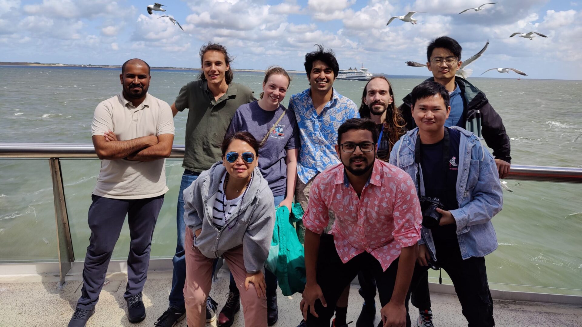
We do interdisciplinary research, combining biology, physics, chemistry, and microfabrication. We extensively use on-chip microfluidic methods and mainly work in a bottom-up fashion. Our research currently covers a wide range of topics, from cytoskeletal dynamics to liquid crystal-based biosensing. We are also keen on diverse collaborative research, ranging from directed evolution to understanding adhesion mechanism of ticks.
Designing biomimetic systems: towards synthetic cells

We are keen on designing and building artificial mimics of natural cells using minimal components, with emphasis on creating out-of-equilibrium conditions. The scope for such an endeavour encompasses both the entities made from and incorporating natural and synthetic components, as well as entities without clearcut biological analogues. A basic starting point for creating synthetic cells is a three-dimensional microcontainer, that separates the self from the surrounding environment. To regulate the thousands of interconnected biochemical reactions, cells have developed various strategies for compartmentalization of their cytoplasm. A relatively recent finding is that of membraneless organelles or biomolecular condensates. Our exploration here is two-fold: (i) To bioengineer membranous vesicles, regulate its morphology, and ultimately make it an active motile system capable of responding to external cues. (ii) Study condensate dynamics within membranous vesicles and carry out distinct biochemical reactions in each of these condensates, thus making rudimentary bioreactors.
Latest publications:
Ganar et al., ACS Synth. Biol., 2022
Ganar et al., COCIS, 2021
Understanding the self-organization in biological systems

We are also using the synthetic cell platform to study the fundamentals of biological systems, especially with regards to the self-assembly of proteins. We are focusing on two processes: (i) studying the actin cytoskeleton that drives shape changes to the cell, and (ii) protein-based condensates that are involved the cytoskeletal functionality as well as potential novel and yet unknown functions. The role of condensates in cytoskeletal functionality and cell shaping is highly unexplored, and we are studying the interplay of condensates and actin dynamics within cell-mimicking vesicles using state-of-the-art microfluidic systems. We frequently collaborate with experts, for example, with Dolf Weijers (WUR) to explore the self-assembly behavior of plant polarity proteins and with Ingrid Dijkgraaf (Maastricht University) to understand the biomechanics of condensate-forming proteins from tick saliva.
Latest publications:
Ganar et al., bioRxiv, 2023
Biosensing using soft matter-based microsensors

Rapid diagnosis is key to ensuring optimized treatment of diseases. Biosensing techniques, however, can be quite slow to perform, often requiring specialized laboratory equipment or technicians to interpret. We want to probe the potential of liquid crystals to exhibit a clear and rapid optical response in presence of biomarkers (antibodies, toxins, etc.). By harnessing their unique ability to exhibit crystalline ordering and birefringence, our goal is to produce a liquid crystal-based biosensor capable of rapid in situ detection of biomolecules. We would ultimately like to prototype a lab-on-a-chip diagnostic test that can be used on-field by a non-expert.
Latest publications:
Honaker et al., J. Mater. Chem. C, 2023
Honaker et al., ACS AMI, 2022
Developing microfluidic technologies

Microfluidic technology offers experimentalists highly controllable and versatile environments. A typical lab-on-a-chip device handles small fluid volumes (in the µL range), flowing through very well-defined channels (of µm dimensions) at low enough flow rates (µL/min or much less). Such setting ensures laminar flow, offering innovative and unique possibilities to control molecules in space and time. Over the years, we have developed numerous microfluidic assays to tackle biological questions: quasi-2D microchambers to study step-by-step biopolymer reactions in a flow-free manner, flow-assays to understand the surface-sensing mechanism in bacteria, physical chemistry-based bubble-blowing machines for making cell-mimicking vesicles, etc. We extensively utilize our on-chip tools, develop them further, and seek to design new ones.
Latest publications:
Chang et al., JoVE, 2023
Last et al., ACS Nano, 2020
Past research
Liposomes as bottom-up cell-mimicking containers

I carried out my postdoctoral research in the lab of Cees Dekker (Delft University of Technology, the Netherlands). With the ambition of creating synthetic cells, we set our primary aim to create and manipulate biochemical containers that could potentially act as architectural scaffolds for artificial cells. I designed a microfluidic technique (Octanol-assisted Liposome Assembly, abbreviated as OLA) to efficiently produce cell-sized and cell membrane-mimicking liposomes. Using OLA, we worked our way towards realising a growth-division cycle of liposomes. We further turned our attention to biomolecular condensates, membraneless organelles crucial for the proper functioning of cells, and succeeded in making ‘condensate-in-liposome’ hybrid containers to obtain a higher level of complexity and biochemical regulation.
Key outcomes:
Deshpande et al., Small, 2019
Deshpande et al., Nat. Commun., 2019
Deshpande et al., Nat. Protoc., 2018
Deshpande et al., ACS Nano, 2018
Deshpande et al., Nat. Commun., 2016
Microfluidics-based single cell/drug-screening assays

During my postdoc, we collaborated with Ulrich Keyser lab (Cambridge University, UK) to further develop the OLA technique as a high-throughput drug-screening platform, for example, to study the effect of peptide antibiotics on membranes. Our assay allows trapping of hundreds of vesicles for long-term experimentation, to quantitatively characterise membranolytic activities, measure permeability coefficients, etc.
During my PhD, we used the flow-free microchambers for developing on-chip assays. For example, we combined them with optical tweezers to develop drug-testing assays on Trypanosoma brucei, a unicellular parasite that causes fatal sleeping sickness in humans. We also teamed up with the lab of Erik van Nimwegen (University of Basel, Switzerland), where we developed an integrated experimental and computational setup to study gene expression at a single-cell level.
Key outcomes:
Schaich et al., Mol. Pharm., 2019
Al Nahas et al., Lab Chip, 2019
Kaiser et al., Nat. Commun., 2018
Surface-sensing mechanism in bacteria

I was a part of a highly interdisciplinary collaboration with the lab of Urs Jenal (University of Basel, Switzerland). We combined molecular biology techniques and mutant studies with microfluidic assays to elucidate the poorly understood surface-sensing mechanism in bacteria. We showed that the flagellar motor acts as a mechanical sensor in Caulobacter crescentus and further identified the key proteins responsible for downstream signalling.
Key outcomes:
Hug et al., Science, 2017
Actin dynamics in quasi-2D confinements

I got exposed to the exciting world of bottom-up biology during my PhD in the lab of Thomas Pfohl (University of Basel, Switzerland), where I explored the dynamics of biopolymer networks. I developed a versatile microfluidic system, termed ‘microchambers’, to achieve step-by-step reaction sequences in a diffusion-controlled manner. I used these microchambers to address important questions regarding the functionality of the actin cytoskeleton, a protein polymer system that helps the cells move and change their shape. I was able to bring to light several emergent properties exhibited by actin bundles and the underlying microstructure dynamics, such as stress built-up in networks, filament length-dependent percolation, etc.
Key outcomes:
Deshpande et al., PLoS ONE, 2015
Deshpande et al., Biomicrofluidics, 2012
